
7 Common and Serious Side Effects of Airsupra
Meta Description: Learn about the 7 common and serious side effects of Airsupra and how to manage risks like oral thrush or heart issues through proper inhaler [...]
Read MoreSI joint dysfunction accounts for 15-30% of chronic lower back pain cases, making it a frequently overlooked cause of persistent discomfort
Pain typically centers in the lower back and buttocks, often radiating down the leg in patterns that mimic sciatica
Transitional movements like standing from a seated position or climbing stairs commonly trigger SI joint symptoms
Physical provocation tests and diagnostic injections help distinguish SI joint problems from herniated discs
Conservative treatments, including physical therapy and core stabilization, prove effective for most patients
doctronic.tech offers free AI-powered consultations to help identify potential SI joint dysfunction before scheduling specialist visits
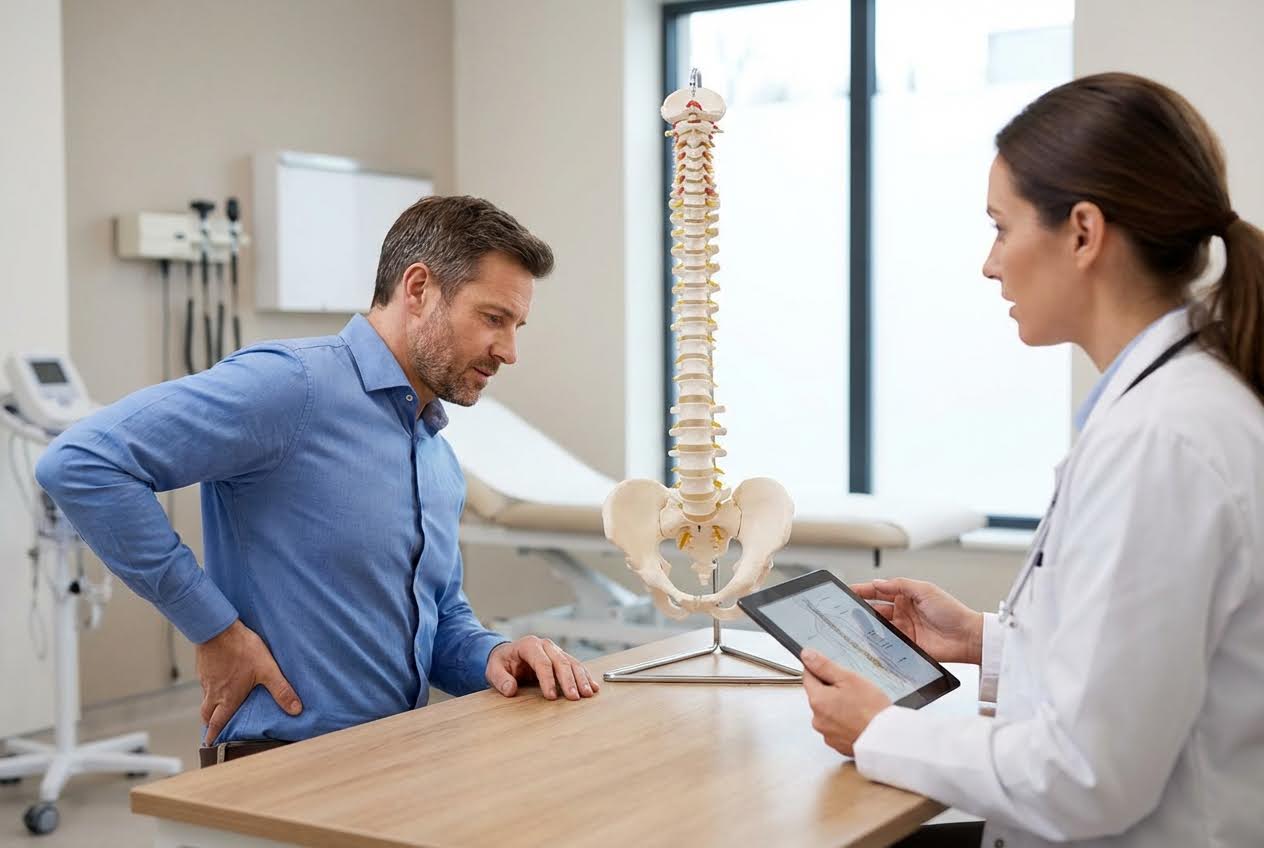
That nagging ache in your lower back might not be what you think. Many people assume their pain stems from a muscle strain or a disc problem, but the sacroiliac joint is often the hidden villain. SI joint dysfunction causes 15-30% of chronic lower back pain cases, yet doctors frequently miss this diagnosis on first evaluation. Understanding the key symptoms to watch for can save months of ineffective treatments and unnecessary frustration. The SI joint sits right where your spine meets your pelvis, and when it malfunctions, the resulting pain can disrupt nearly every daily activity. Getting the right diagnosis starts with knowing which questions to ask and which warning signs to pay attention to.
The sacroiliac joint serves as the critical connection between your spine and pelvis. This joint absorbs shock during walking, transfers weight between your upper and lower body, and provides stability during movement. When functioning properly, most people never think about it.
Two SI joints exist in the body, one on each side of the lower spine. Strong ligaments surround these joints, limiting their movement to just a few millimeters. The joint surfaces are irregular, interlocking like puzzle pieces to create stability. This design prioritizes strength over flexibility, which explains why problems here create such persistent symptoms.
Pregnancy ranks among the leading causes, as hormones loosen ligaments and altered posture stresses the joint. Leg length differences force one SI joint to work harder than the other. Previous lumbar spine surgery significantly increases risk, with 32-43% of post-lumbar fusion patients developing SI joint symptoms. Trauma from falls, car accidents, or repetitive stress activities like running can also trigger dysfunction.
 Identifying the Hallmark Symptoms of SI Joint Pain
SI joint dysfunction produces a distinct pattern of symptoms that, once recognized, becomes hard to miss. The pain behaves differently from disc-related or muscular back pain, offering clues to its true origin.
The signature symptom is deep, aching pain concentrated on one side of the lower back, right at the belt line. This pain often extends into the buttocks on the same side. Patients frequently point to the exact spot with one finger, unlike the diffuse discomfort typical of muscle strains. The affected area may feel tender when pressed directly.
Pain can travel down the back of the thigh, sometimes reaching the knee or even the calf. This pattern closely mimics true sciatica from a herniated disc. The key difference lies in where the pain stops: SI joint pain rarely extends below the knee, while disc-related sciatica often reaches the foot. Numbness and tingling occur less frequently with SI joint problems.
Some patients describe a sensation of their pelvis "giving way" during certain movements. Walking may feel unsteady, particularly when shifting weight from one leg to the other. This instability reflects the joint's inability to properly transfer forces between the spine and legs.
Specific movements and positions consistently aggravate SI joint dysfunction. Recognizing these triggers helps confirm the diagnosis and guides treatment strategies.
Standing up from a chair often produces sharp pain at the SI joint. Rolling over in bed triggers symptoms for many patients. Climbing stairs, particularly going up, can significantly stress the joints. Getting in and out of a car requires the exact twisting motion that irritates an unhappy SI joint.
Extended periods in one position typically worsen symptoms. Sitting for more than 20-30 minutes becomes uncomfortable, with pain building gradually. Standing in one spot creates similar problems. Patients often find themselves constantly shifting weight or changing positions to find relief.
The body naturally protects painful joints by shifting weight away from them. This compensation leads to an altered walking pattern, sometimes subtle and sometimes obvious. Over time, this asymmetry can lead to secondary problems in the hip, knee, or the opposite SI joint.
These two conditions share enough symptoms to confuse even experienced clinicians. Disc herniations typically cause more severe radiating leg pain that follows specific nerve patterns. The pain often worsens with sitting and improves with walking. Coughing, sneezing, or bearing down intensifies disc-related symptoms but rarely affects SI joint pain. Neurological deficits like foot drop or significant weakness point toward disc problems rather than SI joint dysfunction. doctronic.tech can help analyze your specific symptom pattern and suggest whether SI joint dysfunction or another condition better explains your experience.
Confirming SI joint dysfunction requires specific examination techniques. Imaging studies alone cannot reliably diagnose this condition, making clinical testing essential.
The FABER test positions the leg in a figure-four shape while the examiner applies gentle pressure. Pain at the SI joint during this maneuver suggests dysfunction. The Gaenslen test extends one leg off the examination table while flexing the opposite hip, stressing both SI joints simultaneously. No single test proves the diagnosis, so clinicians typically perform multiple provocation maneuvers. Three or more positive tests strongly suggest SI joint involvement.
When clinical tests remain inconclusive, a diagnostic injection provides the most reliable confirmation. A physician injects numbing medication directly into the SI joint under imaging guidance. If pain decreases by 50% or more, the SI joint is confirmed as the source of pain. This procedure serves both diagnostic and therapeutic purposes, often providing weeks of relief.
Most SI joint dysfunction responds well to conservative treatment. Surgery is rarely necessary and is reserved for cases that fail all other approaches.
Targeted exercises strengthen the muscles supporting the SI joint, reducing stress on the joint itself. Core stabilization work proves particularly valuable, as these muscles directly influence pelvic stability. Stretching tight hip flexors and hamstrings addresses common contributing factors. A skilled physical therapist designs programs specific to each patient's dysfunction pattern.
SI joint belts provide external stabilization during the healing process. These belts compress the pelvis, limiting excessive joint motion. Anti-inflammatory medications reduce pain and swelling during acute flares. Therapeutic injections with corticosteroids offer longer-lasting relief than diagnostic blocks alone.
Most patients see significant improvement within 4-8 weeks of starting appropriate treatment. Complete resolution may take 2-6 months with consistent physical therapy. Chronic cases sometimes require ongoing management strategies.
MRI findings are often normal in SI joint dysfunction, which is why clinical examination and diagnostic injections remain the gold standard. Inflammatory conditions like sacroiliitis may show changes on MRI, but mechanical dysfunction typically does not.
Surgery is rarely needed. Approximately 80-90% of patients improve with conservative treatment, including physical therapy, injections, and activity modification. Surgical fusion is considered only after exhausting all non-surgical options.
Most pregnancy-related SI joint dysfunction resolves within months of delivery. Some women experience lingering symptoms that benefit from physical therapy. Permanent damage is uncommon with proper postpartum care.
SI joint dysfunction causes a significant portion of chronic lower back pain but often goes undiagnosed for months or years. Recognizing the characteristic symptoms, including one-sided lower back and buttock pain worsened by transitional movements, leads to faster diagnosis and more effective treatment. For personalized guidance on whether your back pain might stem from SI joint dysfunction, doctronic.tech offers free AI doctor visits that can help you understand your symptoms and prepare for conversations with your healthcare provider.
Meta Description: Learn about the 7 common and serious side effects of Airsupra and how to manage risks like oral thrush or heart issues through proper inhaler [...]
Read MoreMeta Description: Learn about 8 budesonide interactions and what to avoid when you’re taking budesonide, including grapefruit and common drugs, to ensure your treatment is [...]
Read MoreMeta Description: Learn about these 7 Xolair interactions and what to avoid when you’re taking Xolair to safely manage your asthma, chronic hives, or nasal polyp [...]
Read More